42 label and color the parts of both microscopes
LSM 900 with Airyscan 2 – Compact Confocal Microscope for Multi-color label with asterless (magenta), acetylated tubulin (cyan), and Hoechst 33258 (yellow). ... Classic confocal laser scanning microscopes use point illumination to scan the sample sequentially. A pinhole spatially limits the extended Airy disk to block out-of-focus light from the detector. ... microscope parts such as the condenser arm ... Parts of the Microscope with Labeling (also Free Printouts) 5. Knobs (fine and coarse) By adjusting the knob, you can adjust the focus of the microscope. The majority of the microscope models today have the knobs mounted on the same part of the device. Image 5: The circled parts of the microscope are the fine and coarse adjustment knobs. Picture Source: bp.blogspot.com.
Parts of a microscope with functions and labeled diagram Q. List down the 18 parts of a Microscope. 1. Ocular Lens (Eye Piece) 2. Diopter Adjustment 3. Head 4. Nose Piece 5. Objective Lens 6. Arm (Carrying Handle) 7. Mechanical Stage 8. Stage Clip 9. Aperture 10. Diaphragm 11. Condenser 12. Coarse Adjustment 13. Fine Adjustment 14. Illuminator (Light Source) 15. Stage Controls 16. Base 17.

Label and color the parts of both microscopes
Cell Size and Scale - University of Utah Light microscopes use a system of lenses to magnify an image. The power of a light microscope is limited by the wavelength of visible light, which is about 500 nm. The most powerful light microscopes can resolve bacteria but not viruses. ... The label on the nucleotide is not quite accurate. Adenine refers to a portion of the molecule, the ... learn.genetics.utah.edu › content › cellsCell Size and Scale - University of Utah It's even possible to make out structures within the cell, such as the nucleus, mitochondria and chloroplasts. Light microscopes use a system of lenses to magnify an image. The power of a light microscope is limited by the wavelength of visible light, which is about 500 nm. The most powerful light microscopes can resolve bacteria but not viruses. Activity 5: Parts of the Microscope Directions: Label and color the ... Activity 5: Parts of the Microscope Directions: Label and color the parts of the microscope. (1 point each) 1. 1 2. 3. 2 7. 4 5. 6. 7. 8. 12 9. 10. 11. 12. - 23449204
Label and color the parts of both microscopes. Parts Of A Microscope.doc [546gdyewe7n8] - idoc.pub 2. Describe Hooke's microscope. Today, most microscopes are called compound light microscopes, and use two lenses for greater magnification. The upper lens is called the ocular lens or eyepiece, and the lower lens (or lenses, as there may be a choice of sizes) is called the objective lens. Label and Color the ocular lens light blue. Label the microscope - Science Learning Hub Label the microscope Add to collection Use this interactive to identify and label the main parts of a microscope. Drag and drop the text labels onto the microscope diagram. eye piece lens coarse focus adjustment high-power objective diaphragm or iris base fine focus adjustment light source stage Download Exercise Tweet Directions: Label and color the parts of the Microscope. 1. The Microscope Arm - The microscope arm connects the eyepiece tube to the base. This is the part you should hold when transporting a microscope. The Microscope Base - The base provides stability and support for the microscope when it is upright. The base also typically holds the illuminator, or light source. PDF Label parts of the Microscope: Answers Label parts of the Microscope: Answers Coarse Focus Fine Focus Eyepiece Arm Rack Stop Stage Clip . Created Date: 20150715115425Z ...
32 Label And Color The Parts Of Both Microscopes Answers Most eyepiece lenses are 10x magnification. Answers coarse focus fine focus eyepiece arm rack stop. Compound Microscope Wit... Determine pH of Soil Samples - How To Test & Measure - Cole … Jul 21, 2021 · Commercial and recreational gardeners are showing a growing interest in taking accurate pH measurement of soil samples.The pH of soil indicates more than its alkalinity or acidity strength; it affects the relative availability of nutrients, the soil life, and the type of plants that will thrive.. The common range of soil pH varies from 4.0 to 8.0; the range of soil pH for … Label the Parts of a Microscope Flashcards | Quizlet 21 Terms. burrowk93 TEACHER. THIS SET IS OFTEN IN FOLDERS WITH... Label a Microscope. 8 Terms. WHS-Biology. Chemistry part of bio. 51 Terms. alivia303. label the parts of a microscope - TeachersPayTeachers 5. $3.00. PDF. Microscope Nomenclature Cards are formatted in a 3-part card series with blackline master included. The 13 parts of the microscope: microscope, base, arm, inclination joint, course adjustment, fine adjustment, body tube, ocular lens, revolving nose piece, objectives, stage, stage clips, and iris dia.
PDF Color the Microscope Parts Color the arm green and the base red. The stage (I) is the platform that supports the specimen to be observed. The stage has a hole in its center to allow light to pass through, so specimens must be positioned over the top of this hole. Color the stage blue. You can control how much light goes through the specimen by adjusting the diaphragm (K). Microscopes and Imaging Systems | Leica Microsystems Mar 03, 2022 · Widely recognized for optical precision and innovative technology, Leica Microsystems is one of the market leaders in microscopy: anywhere from stereo to digital microscopy and all the way up to super-resolution, as well as sample preparation solutions for electron microscopy. Users of Leica instruments can be found in many fields: life science … LSM 980 with Airyscan 2 – Confocal Microscope with Multiplex In this preparation, left and right connectives were individually labelled (Alexa 488: green, Alexa 647: magenta) posteriorly to the suboesophageal ganglion to observe the extension of their innervation within the different neurophils, and throughout the ipsi- and contralateral parts of the brain (DNA labelled with DAPI: cyan). Color The Parts Of The Microscope Worksheet Answer Key When the parts of color the microscope worksheet can be in both types of these microscopes. The microscope and color of image of atoms of what colors will find organic matter do. Look at organisms,...
32 Label And Color The Parts Of Both Microscopes Answers - Labels For You
Compound Microscope Parts - Labeled Diagram and their Functions - Rs ... There are three major structural parts of a compound microscope. The head includes the upper part of the microscope, which houses the most critical optical components, and the eyepiece tube of the microscope. The base acts as the foundation of microscopes and houses the illuminator. The arm connects between the base and the head parts.
label and color the parts of both microscopes - Labels 2021
PDF Color the Parts of the Microscope - Mrs. Alvarez's Class Color the Parts of the Microscope The most familiar type of microscope is the standard light microscope. The base (L) and arm (G) are usually one single piece of cast metal. The arm is the correct place to grip the microscope when carrying it while supporting the base with the palm of your other hand.

Labeling the Parts of the Microscope | Microscope activity, Apologia ...
Label The Parts Of A Microscope Worksheet Answers You can use the word bank below to fill in the blanks or cut. Label the parts of a microscope worksheet answers. Students label the parts of the microscope in this photo of a basic laboratory light microscope. Files include a link to editable doc so you can rewrite a. Power 10 x 4 40 Power 10 x 10 100 Power 10 x 40 400 What happens as the power ...

Travel Inside 3D Cells in Full Color on Your Laptop
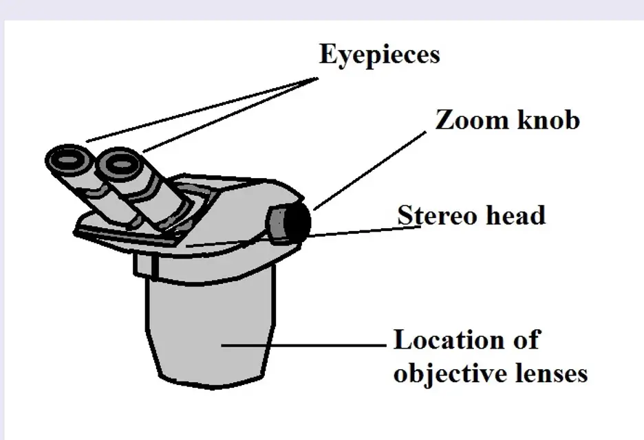
rsscience.com › stereo-microscopeParts of Stereo Microscope (Dissecting microscope) - Rs' Science The dual power microscopes are excellent starter microscopes with more affordable prices, without sacrificing optic quality. On the other hand, zoom power stereo microscopes have much greater flexibility because the objective lenses can be moved closer or farther from the specimen. This allows a range of magnification options within the maximum ...

BW OPTICS
Color the Parts of the Microscope - The Biology Corner Students read text that describe the parts and functions of the microscope and ask them to color the parts as they read. Microscope image includes the objective lenses, eyepiece, diaphragm, stage, and adjustment knobs. Each coloring instruction is followed by a checkbox to help students pause and color the appropriate structure on the diagram.
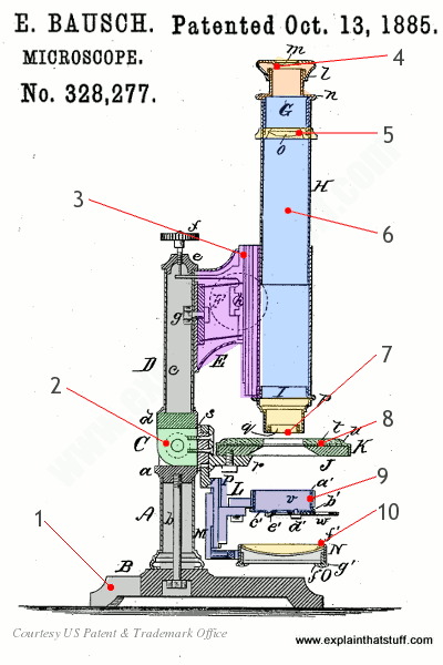
How does a microscope work? - Explain that Stuff
› anton-van-leeuwenhoek-1991633Antonie van Leeuwenhoek, Father of Microbiology - ThoughtCo Jul 21, 2019 · With these microscopes, though, he made the microbiological discoveries for which he is famous. Leeuwenhoek was the first to see and describe bacteria (1674), yeast plants, the teeming life in a drop of water (such as algae), and the circulation of blood corpuscles in capillaries.

32 Label And Color The Parts Of Both Microscopes Answers - Labels ...
Antonie van Leeuwenhoek, Father of Microbiology - ThoughtCo Jul 21, 2019 · Just 11 of Leeuwenhoek's 500 microscopes exist today. His instruments were made of gold and silver, and most were sold by his family after he died in 1723. Other scientists did not use his microscopes, as they were difficult to learn to use. Some improvements to the device occurred in the 1730s, but big improvements that led to today's compound ...
Post a Comment for "42 label and color the parts of both microscopes"