38 diagram of a labeled microscope
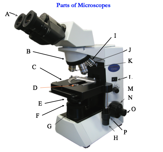
Compound Microscope - Diagram (Parts labelled), Principle and Uses See: Labeled Diagram showing differences between compound and simple microscope parts Structural Components The three structural components include 1. Head This is the upper part of the microscope that houses the optical parts 2. Arm This part connects the head with the base and provides stability to the microscope. Light Microscope- Definition, Principle, Types, Parts, Labeled Diagram ... A light microscope is a biology laboratory instrument or tool, that uses visible light to detect and magnify very small objects and enlarge them. They use lenses to focus light on the specimen, magnifying it thus producing an image. The specimen is normally placed close to the microscopic lens.
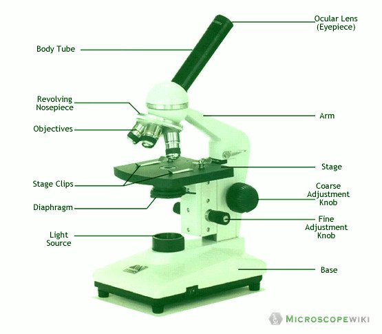
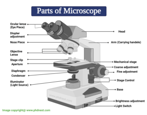
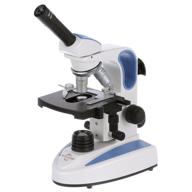
Parts of a microscope with functions and labeled diagram Apr 19, 2022 · Head – This is also known as the body. It carries the optical parts in the upper part of the microscope. Base – It acts as microscopes support. It also carries microscopic illuminators. Arms – This is the part connecting the base and to the head and the eyepiece tube to the base of the microscope.
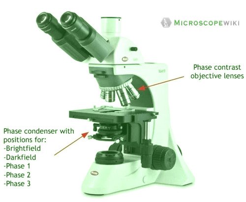
Diagram of a labeled microscope
Pseudostratified Columnar Epithelium under a Microscope with a Labeled ... Pseudostratified Columnar Epithelium under a Microscope with a Labeled Diagram 06/04/2022 by anatomylearner The pseudostratified columnar epithelium comprises a single layer of cells but seems to be multilayered. It is because different cellular heights and nuclei are also placed at a different levels. Simple Microscope - Diagram (Parts labelled), Principle, Formula and Uses Parts of a Simple Microscope A simple microscope consists of Optical parts Mechanical parts Labeled Diagram of simple microscope parts Optical parts The optical parts of a simple microscope include Lens Mirror Eyepiece Lens A simple microscope uses biconvex lens to magnify the image of a specimen under focus. Simple Squamous Epithelium under a Microscope with a Labeled Diagram ... The microscopic image shows a single layer of the flattened nucleus (deep blue). Surrounding the flattened nucleus, you will find pink in color intracellular and extracellular matrix. As there is a single layer of the flattened nucleus that covers the inner surface, it is the simple squamous epithelium.
Diagram of a labeled microscope. Microscope, Microscope Parts, Labeled Diagram, and Functions Microscope, Microscope Parts, Labeled Diagram, and Functions What is Microscope? A microscope is a laboratory instrument used to examine objects that are too small to be seen by the naked eye. It is derived from Ancient Greek words and composed of mikrós, "small" and skopeîn,"to look" or "see". Parts of the Microscope with Labeling (also Free Printouts) Parts of the Microscope with Labeling (also Free Printouts) A microscope is one of the invaluable tools in the laboratory setting. It is used to observe things that cannot be seen by the naked eye. Table of Contents 1. Eyepiece 2. Body tube/Head 3. Turret/Nose piece 4. Objective lenses 5. Knobs (fine and coarse) 6. Stage and stage clips 7. Aperture Testes: Anatomy, definition and diagram | Kenhub Testis. 1/5. The testes (testicles) are male reproductive glands found in a saccular extension of the anterior abdominal wall called the scrotum. They are in ovoid shape, sized four to six centimeters in length. Testes develop retroperitoneally on the posterior abdominal wall and descend to scrotum before birth. Sperm Under Microscope with Labeled Diagram » AnatomyLearner >> The ... Sperm Under Microscope with Labeled Diagram 24/06/202217/06/2022by anatomylearner While studying the histological features of the seminiferous tubules and epididymis, you will see sperm cells under the microscope. They are much smaller and lie in groups along the inner margin of the Sertoli cells.
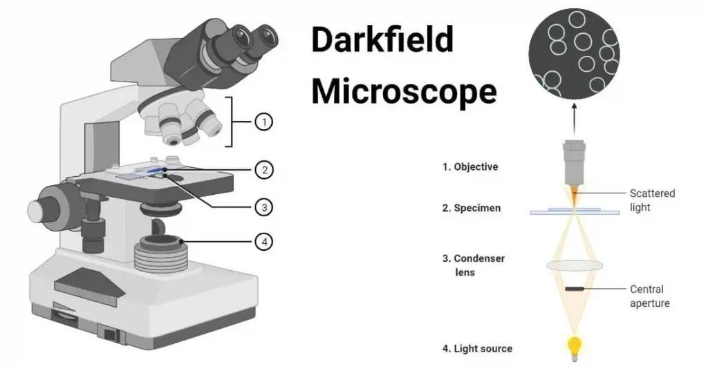
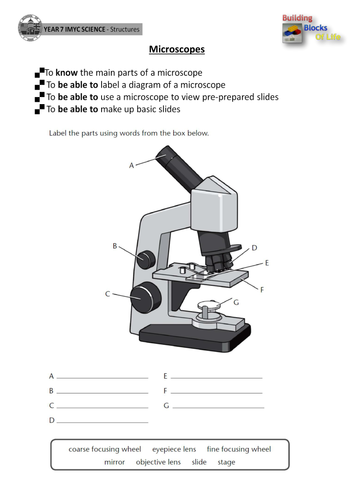
Microscope Diagram Worksheet - The Microscope Create A Labelled Diagram ... Microscope Labeled Diagram from cdn.slidesharecdn.com Used to support the microscope when carried. There is a printable worksheet available for download here so you can take the . This online quiz is called microscope labeling game science, microsope. Be sure to check our teachers notebook store for other printables. Brightfield Microscope (Compound Light Microscope)- Definition ... Parts of a microscope with functions and labeled diagram; 22 Types of Spectroscopy with Definition, Principle, Steps, Uses ... Functions, Labeled Diagram; Disadvantages of Brightfield microscope. The aperture diaphragm may cause great contrast which may distort the outcome of the image, therefore iris diaphragm is preferred. Microscope- Definition, Parts, Functions, Types, Diagram, Uses It is a type of fluorescence microscope that is used to produce 2-D or 3-D images of relatively thick specimens. In this type, the excitation light is focused on a specific spot of sample lying on the focal plane. The focus spot is optically manipulated to scan the entire sample and generate a 3-D image. Electron Microscope Principle, Uses, Types and Images (Labeled Diagram ... Ans: A light microscope has a low resolving power (0.25µm to 0.3µm) while the electron microscope has a resolution power about 250 times higher than the light microscope at about 0.001µm. Similarly, a light microscope has a magnification of 500X to 1500x while the electron microscope has a much higher magnification of 100,000X to 300,000X.
Blood Histology Slides with Description and Labeled Diagram The blood is a specialized connective tissue that is fluid and circulates through the vascular channel. In the blood histology slide, you will find different types of cells with their specific features. This might be a short article where I will show you all the cells from the blood microscope slide with a labeled diagram and actual pictures. Compound Light Microscope Diagram Worksheet - Google Groups Study manual following chapter which describes features of the initial light microscope and the function of each carbon the diagram of the microscope below. You will label sketches to compound light microscope worksheet may want to your students to use worksheets to. On a typical student compound light microscope there are 3-4 of objective lenses. Labeled diagram of an atom (Including History and ... - The Boffins Portal Labeled diagram of an atom. If you are looking for a labeled diagram of an atom, here it is: Source: 1, 2. ... Scientists in the earlier years found that they could never peep into the insides of an atom using the microscope to know its structure. It was too small for a microscope to be seen. So instead, scientists used clever guesses (called ... Microscope Types (with labeled diagrams) and Functions Simple microscope labeled diagram Simple microscope functions It is used in industrial applications like: Watchmakers to assemble watches Cloth industry to count the number of threads or fibers in a cloth Jewelers to examine the finer parts of jewelry Miniature artists to examine and build their work Also used to inspect finer details on products
Microscope Types (with labeled diagrams) and Functions
Simple Microscope - Parts, Functions, Diagram and Labelling Simple Microscope - Parts, Functions, Diagram and Labelling A microscope is one of the commonly used equipment in a laboratory setting. A microscope is an optical instrument used to magnify an image of a tiny object; objects that are not visible to the human eyes. Table of Contents The common types of microscopes are: What is a Simple microscope?

Compound Microscope – Diagram (Parts labelled), Principle and ...
› 6-label-the-microscopeLabel the microscope — Science Learning Hub Jun 08, 2018 · Use this interactive to identify and label the main parts of a microscope. Drag and drop the ...
Microscope Types (with labeled diagrams) and Functions
Spectroscope Diagram, Parts & Function - Study.com The spectroscope diagram shows what happens to white light when it shines through a spectroscope. The spectroscope has multiple parts that work together to produce a spectrum.
Bright Field Microscope: Definition, Parts, Diagram ...
Plant Cell Under Microscope Labeled 40X : Young Root 2 Of Broad Bean ... A cell is a very tiny structure which exists in living bodies. Which part is not visible when looking at plant and animal cells under a microscope. Label the diagram of the following plant cell. Microscope Magnification Ppt Download from slideplayer.com Chloroplast, nucleus, cytoplasm, cytoplasm, vacuole, cell membrane 9. (iv) describe how you ...
Vektor Stok Microscope Diagram Vector Illustration Labeled ...
Draw a diagram of a matured microspore of an angiosperm. Label its ... Draw a diagram of L.S. of an anatropous ovule of an angiosperm and label the following parts. asked Aug 17, 2021 in Biology by Devakumari ( 52.3k points) sexual reproduction in flowering plants
Microscope- Definition, Parts, Functions, Types, Diagram, Uses
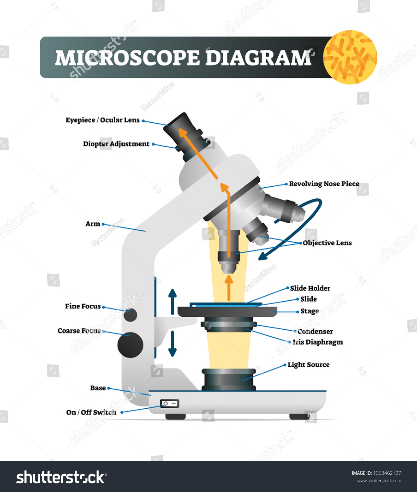
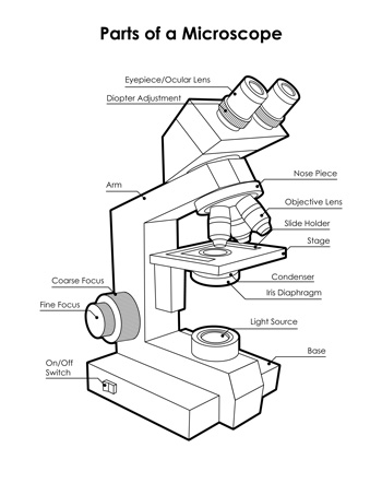
Compound Microscope- Definition, Labeled Diagram, Principle, Parts, Uses A beam of visible light from the base is focused by a condenser lens onto the specimen. The objective lens picks up the light transmitted by the specimen and creates a magnified image of the specimen called the primary image inside the body tube. This image is again magnified by the ocular lens or eyepiece.

Parts of Microscope, Function, Names & Labeled Diagram ...
Neuron under Microscope with Labeled Diagram - AnatomyLearner But, first, let's try to identify the following features from a neuron with the help of a labelled diagram. Cell body or perikaryon of a neuron Nucleus, cytoplasm, the plasma membrane of a neuron Nissl bodies in the cell body of a neuron An initial segment of axon and axon hillock Dendrites and axons of a neuron Axolemma and myelin sheath

Labelling a Microscope Diagram | Quizlet
Inverted Microscope- Definition, Principle, Parts, Labeled Diagram ... The specimen is placed on a large stage that can be able to hold. With the objectives located below the stage and pointing upwards, it collects light from the condenser magnifying the image, which is then sent to the ocular lens. Light is reflected by the ocular lens through a mirror.

Simple Microscope - Diagram (Parts labelled), Principle ...
Bright-field microscope (Compound light microscope) - Diagram (Parts ... Bright-field microscope parts (Labeled Diagram) Ocular Lens This microscope has two eye lenses or ocular lens on the top of the microscope that are used to focus the image from the objective lens. It is from these lenses that we see the magnified image of the specimen. Objective Lens

Parts of Microscope, Function, Names & Labeled Diagram ...
Electron Microscope-Definition, Principle, Types, Uses, Labeled Diagram Parts of an Electron Microscope The electron microscope is placed vertically and has the shape of a tall vacuum column. It consists of the following elements: 1. Electron gun A heated tungsten filament that produces electrons makes up the electron cannon. 2. Electromagnetic lenses The condenser lens directs the electron beam to the specimen.

Microscope With Labels Clip Art at Clker.com - vector clip ...
rsscience.com › compound-microscope-parts-labeledCompound Microscope Parts – Labeled Diagram and their ... There are two major optical lens parts of a microscope: Eyepiece (10x) and Objective lenses (4x, 10x, 40x, 100x). Total magnification power is calculated by multiplying the magnification of the eyepiece and objective lens. The illuminator provides a source of light. The light is focused by the condenser and passing through the specimen placed ...
Label a microscope - Teaching resources
Microscope: Types of Microscope, Parts, Uses, Diagram - Embibe There microscope anatomy includes three structural parts, i.e. head, base, and arm. Head - This is also known as the body; it carries the optical parts in the upper part of the microscope.. Base - It acts as microscopes support.It also carries microscopic illuminators. Arms - The microscope arm connects the base and the head and the eyepiece tube to the microscope base.
Microscope - Teaching resources
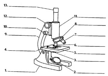
Microscope Parts, Function, & Labeled Diagram - slidingmotion Microscope parts labeled diagram gives us all the information about its parts and their position in the microscope. Microscope Parts Labeled Diagram The principle of the Microscope gives you an exact reason to use it. It works on the 3 principles. Magnification Resolving Power Numerical Aperture. Parts of Microscope Head Base Arm Eyepiece Lens

Microscope Labeling #1 Diagram | Quizlet
Simple Squamous Epithelium under a Microscope with a Labeled Diagram ... The microscopic image shows a single layer of the flattened nucleus (deep blue). Surrounding the flattened nucleus, you will find pink in color intracellular and extracellular matrix. As there is a single layer of the flattened nucleus that covers the inner surface, it is the simple squamous epithelium.

Microscope Diagram - Label Diagram | Quizlet
Simple Microscope - Diagram (Parts labelled), Principle, Formula and Uses Parts of a Simple Microscope A simple microscope consists of Optical parts Mechanical parts Labeled Diagram of simple microscope parts Optical parts The optical parts of a simple microscope include Lens Mirror Eyepiece Lens A simple microscope uses biconvex lens to magnify the image of a specimen under focus.

Microscope Labeling Diagram | Quizlet
Pseudostratified Columnar Epithelium under a Microscope with a Labeled ... Pseudostratified Columnar Epithelium under a Microscope with a Labeled Diagram 06/04/2022 by anatomylearner The pseudostratified columnar epithelium comprises a single layer of cells but seems to be multilayered. It is because different cellular heights and nuclei are also placed at a different levels.
Compound Microscope Parts, Functions, and Labeled Diagram ...

Parts of a Microscope with Their Functions • Microbe Online

Pic Of Microscope

Left Side of Microscope Labeling Diagram | Quizlet

Compound Microscope- Definition, Labeled Diagram, Principle ...

Compound Microscope Parts – Labeled Diagram and their ...

2.1 " Compound Microscope" | Download Scientific Diagram

Types of Microscopes: Definition, Working Principle, Diagram ...
Label microscope - Teaching resources
Labeling Microscope Worksheet | Teaching Resources

Light Microscope- Definition, Principle, Types, Parts ...
biology labeled microscope diagram - Clip Art Library

Microscope Terms Glossary | Earth science lessons, Medical ...
Experiment 1B | Lab01 | Virtual Edge| General Microbiology ...

File:Microscope diagram.png - Wikimedia Commons
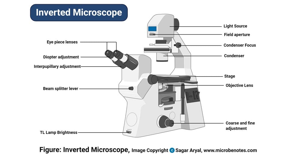
Inverted Microscope- Definition, Principle, Parts, Labeled ...
Compound Microscope Parts, Functions, and Labeled Diagram ...
Microscope Diagram Labeled, Unlabeled and Blank | Parts of a ...
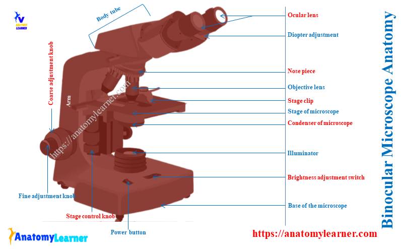
Binocular Microscope Anatomy - Parts and Functions with a ...

How to draw compound of Microscope easily - step by step
Microscope Parts and Function

Compound microscope labeling Diagram | Quizlet
Post a Comment for "38 diagram of a labeled microscope"