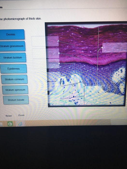
40 label the photomicrograph of the skin and its accessory structures.
Answered: 1. In the photomicrograph below of… | bartleby 1. In the photomicrograph below of cartilage tissue, find and label the indicated structures. Extra cellul (ar m Lacuna Chondrocyte droyte Elastic protein fibers Extracellular matrix 2. In the photomicrograph below of compact bone tissue, find and label the indicated structu p Osteon Lamella Lacuna o Osteocyte Canaliculi » Central canal ... Skin Anatomy: The Layers of Skin and Their Functions The skin is the body's largest organ. It is made of three layers, each of which has specific functions. The outermost epidermis is responsible for producing new skin cells, protecting the body from unwanted substances, and retaining moisture to keep the skin well hydrated. The middle dermis is responsible for supporting and strengthening the skin.
Structure and Function of Skin | Biology for Majors II | | Course Hero The skin and its accessory structures make up the integumentary system, which provides the body with overall protection. The skin is made of multiple layers of cells and tissues, which are held to underlying structures by connective tissue (Figure 1). The deeper layer of skin is well vascularized (has numerous blood vessels).

Label the photomicrograph of the skin and its accessory structures.
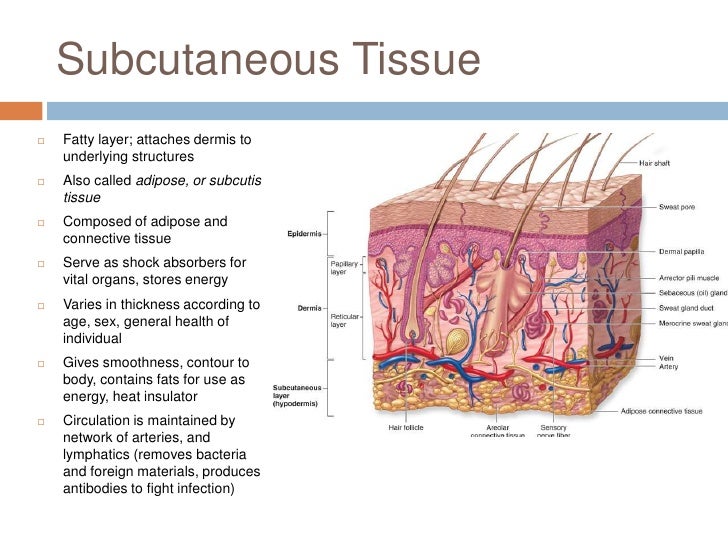
Solved Label the photomicrograph of the skin and its Question: Label the photomicrograph of the skin and its accessory structures. Sebaceous gland Duct of sebaceous gland Epidermis Hair follicle.1 answer · Top answer: Please find the labeled picture attached and also the explanation of the terms below: Sebaceous gland: Sebaceous gland lubricate... Accessory Structures of the Skin - Anatomy & Physiology Accessory structures of the skin include hair, nails, sweat glands, and sebaceous glands. These structures embryologically originate from the epidermis and can extend down through the dermis into the hypodermis. Hair Hair is a keratinous filament growing out of the epidermis. It is primarily made of dead, keratinized cells. Label The Photomicrograph Of The Sebaceous Gland - Era Elang Accessory structures of the skin include hair, nails, sweat glands, and sebaceous glands. Before placing your slide on the microscope stage, remember to read the label, examine the slide with your eye and note any visible macroscopic features that . Hair sebaceous gland dermis hair follicle epidermis duct of sebaceous.
Label the photomicrograph of the skin and its accessory structures.. Accessory Structures of the Skin | Anatomy and Physiology I | | Course Hero Accessory structures of the skin include hair, nails, sweat glands, and sebaceous glands. These structures embryologically originate from the epidermis and can extend down through the dermis into the hypodermis. Hair Figure 1. Hair follicles originate in the epidermis and have many different parts. Solved Label the photomicrograph of the skin and its | Chegg.com See the answer Label the photomicrograph of the skin and its accessory structures Epidermis Duct of sebaceous gland Hair follicle Sebaceous gland Show transcribed image text Expert Answer 2. The picture here demonstrates the pseudostratified columnar epithelium. 5.1 Layers of the Skin - Anatomy & Physiology Accessory structures, hair, glands, and nails, are found associated with the skin. The deeper layer of skin is well vascularized (has numerous blood vessels) and is superficial to the hypodermics. It also has numerous sensory, and autonomic and sympathetic nerve fibers ensuring communication to and from the brain. PDF The Integumentary System - Holly H. Nash-Rule, PhD Label the skin structures and areas indicated in the accompanying diagram of thin skin. Then, complete the statements that ... Accessory Organs of the Skin 9. Match the key choices with the appropriate descriptions. Some terms are used more than once. Key: a. arrector pili d. hair follicle g.
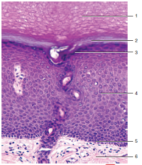
5.2 Accessory Structures of the Skin - Anatomy & Physiology Accessory structures of the skin include hair, nails, sweat glands, and sebaceous glands. These structures embryologically originate from the epidermis and can extend down through the dermis into the hypodermis. Hair Hair is a keratinous filament growing out of the epidermis. It is primarily made of dead, keratinized cells. Label The Photomicrograph Of The Skin And Its Accessory Structures ... The Integumentary System A. Anatomy of the skin. Identify and label the photograph of the skin model: epidermis, dermis, dermal papillae, hypodermis, hair follicle, hair bulb, sebaceous gland, sudoriferous gland, duct of sudoriferous gland, pore of... Posted 2 months ago Q: Diagram of the skin and accessory structures - Quizlet Diagram of the skin and accessory structures STUDY Flashcards Learn Write Spell Test PLAY Match Gravity Hair shaft Click card to see definition 👆 ... Click again to see term 👆 1/9 Created by jcgrainson Terms in this set (9) Hair shaft Hair root Sebaceous gland Arrector pili muscle Hair follicle Hair bulb Eccrine sweat gland Papilla of hair PDF Name the Condition - Dr. Scott Croes' Website the cartoon and the photomicrograph. •Name the Layers of skin and label the dermal papilla and dermis •Name the Layers of skin and label the dermal papilla and dermis. Name the layer of skin shown. Stratum Spinosum. Name the specific layers of skin indicated by the ... Identify the following structures: Epidermis, Hair cortex, Hair medulla ...
Solved Label the Photomicrograph of the skin and its - Chegg Who are the experts? Experts are tested by Chegg as specialists in their subject area. We review their content and use your feedback to keep the quality high. Transcribed image text: Label the Photomicrograph of the skin and its accessory structures. Figure 7.1: Photomicrograph of Skin Diagram - Quizlet Start studying Figure 7.1: Photomicrograph of Skin. Learn vocabulary, terms, and more with flashcards, games, and other study tools. Diagram of human skin structure - Science Learning Hub Rights: University of Waikato Published 1 February 2011 Size: 100 KB Referencing Hub media The epidermis is a tough coating formed from overlapping layers of dead skin cells. Appears in ARTICLE Touch Our sense of touch allows us to receive information about our internal and external environments, making it important for sensory perception. Function And Structure of Skin And Subcutaneous Tissue - Earth's Lab Structure of The Skin And Subcutaneous Tissue. The skin is thickest in areas subjected to wear and tear (abrasion), such as the soles of the feet, where it may be 6 mm in thickness. It is thinnest on the eyelids, eardrums, and external genitalia, where it averages about 0.5 mm in thickness. The skin consists of two major layers: the epidermis ...
Accessory Structures of the Skin - YouTube
Solved Label the photomicrograph of the skin and its | Chegg.com Label the photomicrograph of the skin and its accessory structures. Epidermis Sebaceous gland Hair follicle Duct of sebaceous gland Label the photomicrograph of the skin and its accessory structures. Epidermis Sebaceous gland Hair follicle Duct of sebaceous gland ; Question: Label the photomicrograph of the skin and its accessory structures ...
Biology Archive | March 09, 2017 | Chegg.com
PreLab03a Integument & Prelab03b Integument Histology Flashcards - Quizlet composed of 5 layers is avascular composed of keratinocytes most superficial layer Middle layer: composed of 2 layers contains hair follicles contains sweat glands Bottom layer: also known as subcutaneous composed of loose areolar and adipose tissue contains sweat glands Place the following layers in order from superficial too deep. epidermis

Review for Cell Bio Final - Slides
Laboratory Manual for Anatomy and Physiology Connie Allen, Valerie Harper · 2020 · ScienceThe accessory structures of the skin include hairs, hair follicles, nails, ... from its normal angle to a 90° angle (perpendicular) with the skin surface, ...
Print Anatomy Exam 1 flashcards | Easy Notecards
Integumentary System HW_answers.docx - Course Hero Label the structures of the fingernail in a lateral view. 14. ... Label the photomicrograph of the skin and its accessory structures. 19. Label the photomicrograph of the sebaceous gland. 20. Label the structures of merocrine sweat glands. 21. Label the structures of the hair follicle. 22.
Anatomy Of The Skin Lecture
Layers of the Skin - Anatomy and Physiology The skin and its accessory structures make up the integumentary system, which provides the body with overall protection. The skin is made of multiple layers of cells and tissues, which are held to underlying structures by connective tissue (). The deeper layer of skin is well vascularized (has numerous blood vessels).
35 Label The Photomicrograph Of Thick Skin - Label Design Ideas 2020
Skin 2: accessory structures of the skin and their functions All are important in the skin's key functions, including protection, thermoregulation and its sensory roles. This article, the second in a two-part series, looks at the structure and function of the main accessory structures of the skin. Citation: Lawton S (2020) Skin 2: accessory structures of the skin and their functions.
Post a Comment for "40 label the photomicrograph of the skin and its accessory structures."