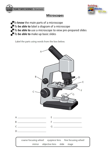
45 label microscope diagram
Animal Cell Electron Microscope Labelled - Q14 Draw a large diagram of ... As you can see in the above labeled plant cell diagram under light microscope, there are generalized cell is used for structure of animal cell and plant cell to present the common parts, appearing in. Plant and animal cells have a nucleus inside the cytoplasm. With the invention of the electron microscope a whole new world was open up to ... Draw And Label Diagram Of Animal Cell - Adolfo Bridgman Check this diagram and learn m. The first is a colored and labeled cell diagram. There are six animal cell diagrams to choose from. Animal cell electron micrograph labelling. 2.3.1 draw and label a diagram of the ultrastructure of a liver cell as an example of an animal cell. The working together of all cells gives an animal its ability.
Compound Microscope- Definition, Labeled Diagram, Principle, Parts, Uses The naked eye can now view the specimen at magnification 400 times greater and so microscopic details are revealed. Alternatively, the magnification of the compound microscope is given by: m = D/ fo * L/fe where, D = Least distance of distinct vision (25 cm) L = Length of the microscope tube fo = Focal length of the objective lens

Label microscope diagram
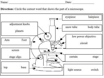
Label The Parts Of A Microscope Worksheet Answers Below you will find both the label the microscope activity worksheet as well as one with answers. Microscope Parts and Use Worksheet Answer Key Along with Labeling the Parts Of the Microscope Blank Diagram Available for. Coarse adjustment knob 13. Once you find your worksheet s you can either click on the pop-out icon or download button to print. Microscope Types (with labeled diagrams) and Functions Simple microscope labeled diagram Simple microscope functions It is used in industrial applications like: Watchmakers to assemble watches Cloth industry to count the number of threads or fibers in a cloth Jewelers to examine the finer parts of jewelry Miniature artists to examine and build their work Also used to inspect finer details on products Stage Of Mitosis Blank Worksheet - 18 images - label microscope diagram ... [Stage Of Mitosis Blank Worksheet] - 18 images - mitosis vs meiosis worksheet answers promotiontablecovers, mitosis worksheet diagram identification, bro dimas, mitosis sequencing worksheet preschool worksheet gallery,
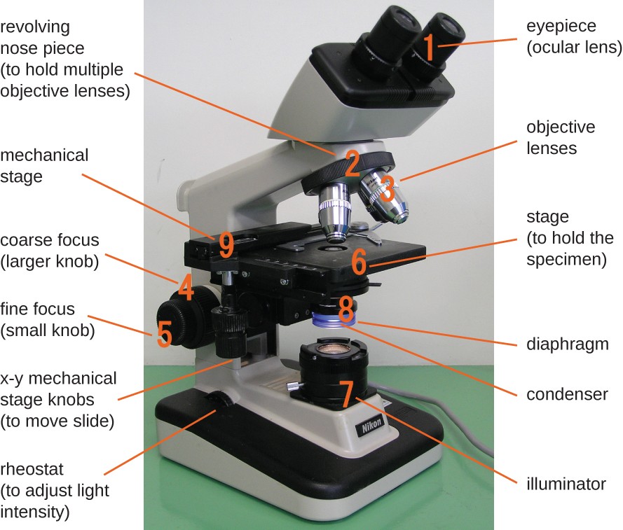
Label microscope diagram. Binocular Microscope Anatomy - Parts and Functions with a Labeled Diagram Now, I will discuss the details anatomy of the light compound microscope with the labeled diagram. Why it is called binocular: because it has two ocular lenses or an eyepiece on the head that attaches to the objective lens, this ocular lens magnifies the image produced by the objective lens. Binocular microscope parts and functions Spectroscope Diagram, Parts, & Function - Study.com The spectroscope diagram shows what happens to white light when it shines through a spectroscope. The spectroscope has multiple parts that work together to produce a spectrum. Plant Cell Under Microscope Labeled 40X - Sadie Bermingham A cell is a very tiny structure which exists in living bodies. Which part is not visible when looking at plant and animal cells under a microscope. Label the diagram of the following plant cell. Microscope Magnification Ppt Download from slideplayer.com Chloroplast, nucleus, cytoplasm, cytoplasm, vacuole, cell membrane 9. (iv) describe how you ... Microscope, Microscope Parts, Labeled Diagram, and Functions Microscope, Microscope Parts, Labeled Diagram, and Functions What is Microscope? A microscope is a laboratory instrument used to examine objects that are too small to be seen by the naked eye. It is derived from Ancient Greek words and composed of mikrós, "small" and skopeîn,"to look" or "see".
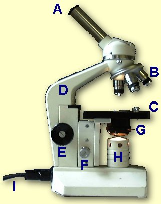
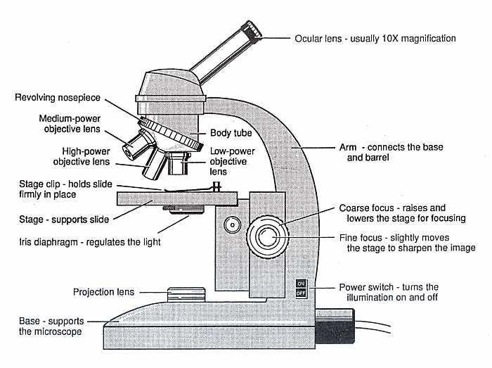
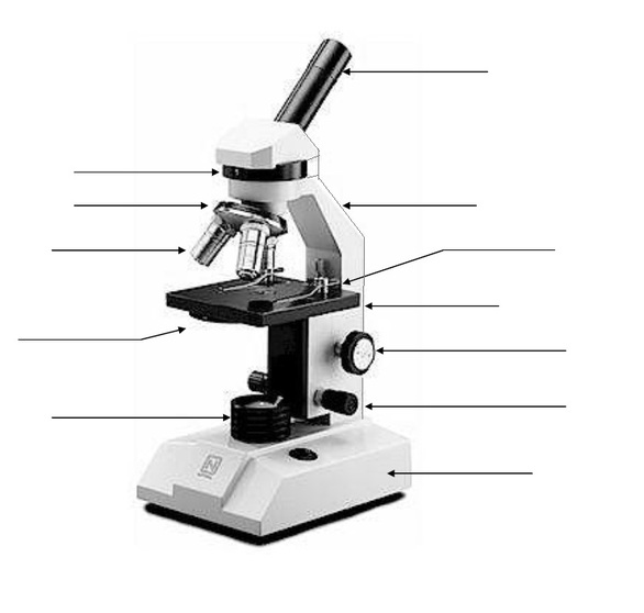
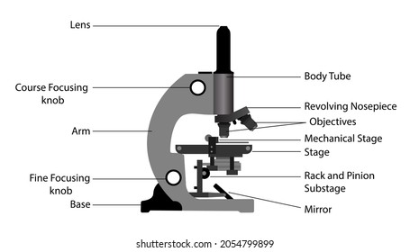
Microscope Quiz: How Much You Know About Microscope Parts ... - ProProfs Projects light upwards through the diaphragm, the specimen, and the lenses. 5. Is used to regulates the amount of light on the specimen. Supports the slide being viewed. Moves the stage up and down for focusing. 6. Is used to support the microscope when carried. Moves the stage slightly to sharpen the image. Draw a diagram of a matured microspore of an angiosperm. Label its ... Draw a diagram of L.S. of an anatropous ovule of an angiosperm and label the following parts. asked Aug 17, 2021 in Biology by Devakumari ( 52.3k points) sexual reproduction in flowering plants Microscopy- History, Classification, Terms, Diagram - The Biology Notes A simple or compound light microscope is used in this technique. It uses transmitted visible light to develop magnified images. It has a low contrasting capacity, low optical resolution, requires staining and has a limited magnification of around 1300X. It is simple and can be used to observe living cells and microorganisms. 2 Dark Field Microscopy Parts of a microscope with functions and labeled diagram - Microbe Notes Figure: Diagram of parts of a microscope There are three structural parts of the microscope i.e. head, base, and arm. Head - This is also known as the body. It carries the optical parts in the upper part of the microscope. Base - It acts as microscopes support. It also carries microscopic illuminators.
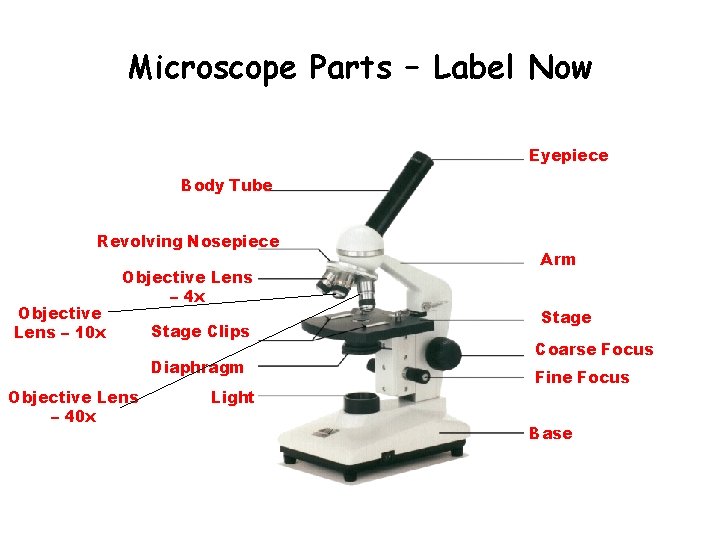
Parts of the Microscope with Labeling (also Free Printouts) Parts of the Microscope with Labeling (also Free Printouts) By Editorial Team March 7, 2022 A microscope is one of the invaluable tools in the laboratory setting. It is used to observe things that cannot be seen by the naked eye. Table of Contents 1. Eyepiece 2. Body tube/Head 3. Turret/Nose piece 4. Objective lenses 5. Knobs (fine and coarse) 6. Testes: Anatomy, definition and diagram | Kenhub Put your knowledge to the test with our reproductive system labeling diagrams and quizzes. Each of the 200-300 lobules of the testis are filled with one to four highly convoluted seminiferous tubules which each course towards the mediastinum testis. Light Microscope (Theory) - Amrita Vishwa Vidyapeetham Microscope can be separated into optical theory microscopes (Light microscope), electron microscopes (eg.TEM, SEM) and scanning probe microscopes. (eg.AFM, PSTM). Optical microscopes function on the basis of optical theory of lenses by which it can magnifies the image obtained by the movement of a wave through the sample. The waves used in ... Simple Microscope - Diagram (Parts labelled), Principle, Formula and Uses Parts of a Simple Microscope A simple microscope consists of Optical parts Mechanical parts Labeled Diagram of simple microscope parts Optical parts The optical parts of a simple microscope include Lens Mirror Eyepiece Lens A simple microscope uses biconvex lens to magnify the image of a specimen under focus.
Microscope labeled diagram
Electron Microscope-Definition, Principle, Types, Uses, Labeled Diagram Parts of an Electron Microscope The electron microscope is placed vertically and has the shape of a tall vacuum column. It consists of the following elements: 1. Electron gun A heated tungsten filament that produces electrons makes up the electron cannon. 2. Electromagnetic lenses The condenser lens directs the electron beam to the specimen.

File:Labelledmicroscope.gif - Wikimedia Commons
Sperm Under Microscope with Labeled Diagram - AnatomyLearner Sperm Under Microscope 400X Labeled Diagram Before that, you may also read the below-mentioned article to get a full idea of the structure of seminiferous tubules - Histological features of the seminiferous tubules with the labeled diagram Okay, first, let's see the different histological features of the seminiferous tubules of an animal.
Microscope
Parts of Telescope, Names, Functions & Diagram - slidingmotion Microscope Parts, Function, & Labeled Diagram. Parts of Computer, Names, Functions & Diagram. Hydraulic Cylinder Parts, Names & Diagrams. Post navigation. Previous Previous post: Parts of Ship, Names, Functions & Diagram. Next Next post: What are the Types of Loads. Leave a Reply Cancel reply. Search. Recent Posts.

Microscope Labeling Diagram | Quizlet
Microscope Diagram Worksheet - The Microscope Create A Labelled Diagram ... This online quiz is called microscope labeling game science, microsope. When you can identify a part of the microscope place the . Using the terms listed below, label the microscope diagram. Use the words from this word list to identify the parts of the microscope. Can be rotated to change magnification.

ABOUT MICROSCOPES | Scienceart
Parts of a Microscope: Lesson for Kids - Study.com Slide your finger down to the base of the microscope and look for a wheel that you can turn. This controls the light intensity coming through the image. Turn that wheel while looking through the...
Lab - Microscope: MAH-Summer 2019-Anatomy and Physiology I
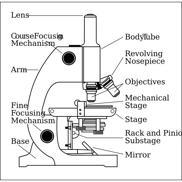
Microscope Parts, Function, & Labeled Diagram - slidingmotion Microscope parts labeled diagram gives us all the information about its parts and their position in the microscope. Microscope Parts Labeled Diagram The principle of the Microscope gives you an exact reason to use it. It works on the 3 principles. Magnification Resolving Power Numerical Aperture. Parts of Microscope Head Base Arm Eyepiece Lens

Microscope parts 3D learning for Android - APK Download
Microscope Diagram - cell division of e coli with continuous media flow ... Microscope Diagram - 15 images - give a well labelled diagram of compound microscope using of typical, bio tem biological transmission electron microscope university, labelled microscope diagram gcse micropedia, a compound microscope diagram micropedia,
Print Map Quiz: Labeling the Microscope ()
Simple Microscope - Parts, Functions, Diagram and Labelling Simple Microscope - Parts, Functions, Diagram and Labelling By Editorial Team March 7, 2022 A microscope is one of the commonly used equipment in a laboratory setting. A microscope is an optical instrument used to magnify an image of a tiny object; objects that are not visible to the human eyes. Table of Contents

Produk Microscope | UD Berkah Abadi
Microscope: Parts Of A Microscope With Functions And Labeled Diagram. Figure: A diagram of a microscope's components. The microscope has three basic components: the head, the base, and the arm. Head:Occasionally, the head is considered the body. It holds the optical components of the upper part of the microscope. Base:The microscope's base provides great support.
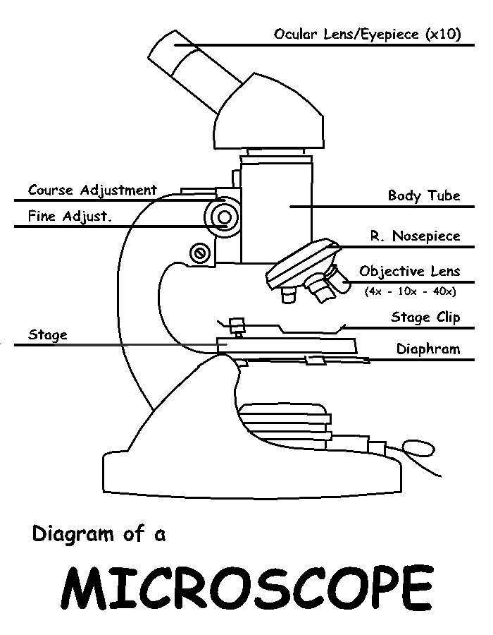
Diagram of a Microscope by ScienceDoodles on DeviantArt
Microscope: Types of Microscope, Parts, Uses, Diagram - Embibe There microscope anatomy includes three structural parts, i.e. head, base, and arm. Head - This is also known as the body; it carries the optical parts in the upper part of the microscope.. Base - It acts as microscopes support.It also carries microscopic illuminators. Arms - The microscope arm connects the base and the head and the eyepiece tube to the microscope base.

Parts of a Microscope Labeling Activity
Oscillatoria | The Blue Green Algae (Guide 2022) - Botnam If fresh material is observed under the microscope specific oscillating movement is observed. Oscillatoria Labeled Diagram. Oscillatoria Structure: A, Few filaments; B, Single enlarged filament; C, a single cell. Oscillatoria Cell Structure. All cells have a well-developed cell wall. The cell wall consists of an inner thin cellular layer a ...

Compound Microscope – Diagram (Parts labelled), Principle and ...
Stage Of Mitosis Blank Worksheet - 18 images - label microscope diagram ... [Stage Of Mitosis Blank Worksheet] - 18 images - mitosis vs meiosis worksheet answers promotiontablecovers, mitosis worksheet diagram identification, bro dimas, mitosis sequencing worksheet preschool worksheet gallery,

This is a common compound microscope Label its parts class 11 ...
Microscope Types (with labeled diagrams) and Functions Simple microscope labeled diagram Simple microscope functions It is used in industrial applications like: Watchmakers to assemble watches Cloth industry to count the number of threads or fibers in a cloth Jewelers to examine the finer parts of jewelry Miniature artists to examine and build their work Also used to inspect finer details on products

Biology label part of microscope
Label The Parts Of A Microscope Worksheet Answers Below you will find both the label the microscope activity worksheet as well as one with answers. Microscope Parts and Use Worksheet Answer Key Along with Labeling the Parts Of the Microscope Blank Diagram Available for. Coarse adjustment knob 13. Once you find your worksheet s you can either click on the pop-out icon or download button to print.
Label microscope - Teaching resources
Microscope side vector drawing with parts labelled | Free SVG

Parts of the Microscope with Labeling (also Free Printouts ...

MICROSCOPE Labeling - Part - 3

Label the Microscope Diagram | Download Scientific Diagram

Parts of a Microscope with Their Functions • Microbe Online

How to Draw a Microscope and Label Its Parts

Compound Microscope: Parts of Compound Microscope
Labeling Microscope Worksheet | Teaching Resources
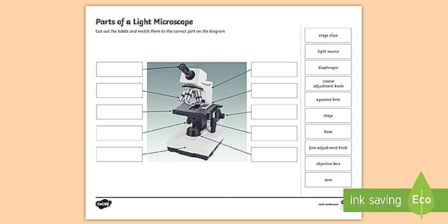
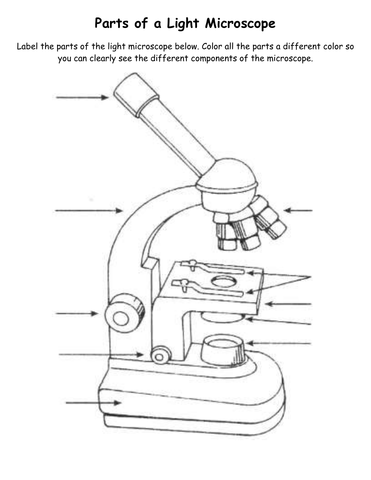
Parts of a Light Microscope Activity | Labeling Task
The Compound Light Microscope Label the following parts on ...

Label the light microscope | Teaching Resources
Free Microscope Drawing, Download Free Microscope Drawing png ...
Instruments of Microscopy | Microbiology | | Course Hero
Scientific Tools Microscope Birth of the Microscope 1590

This is a common compound microscope. Label its parts from A ...
Label Parts Of Microscope - ClipArt Best

Microscope World Blog: Labeling the Parts of the Microscope

Microscope exercise for Grade 3

Parts of a Microscope Microscope Basics. Label the Compound ...

Draw a labelled diagram of a compound microscope.

Microscope Diagram Labeled, Unlabeled and Blank | Parts of a ...

Diagram of diatom microscope slide positioned with its label ...

Microscope With Labels Clip Art at Clker.com - vector clip ...

Cytology. Cytology. radiation used to illuminate the specimen ...

Labeling a Microscope Free Worksheet Pack
Vektor Stok Different Parts Microscope (Tanpa Royalti ...
Modified Science Diagram; Label Parts of a Microscope; Special Education

Label Microscope Parts - ClipArt Best
Parts of a Microscope
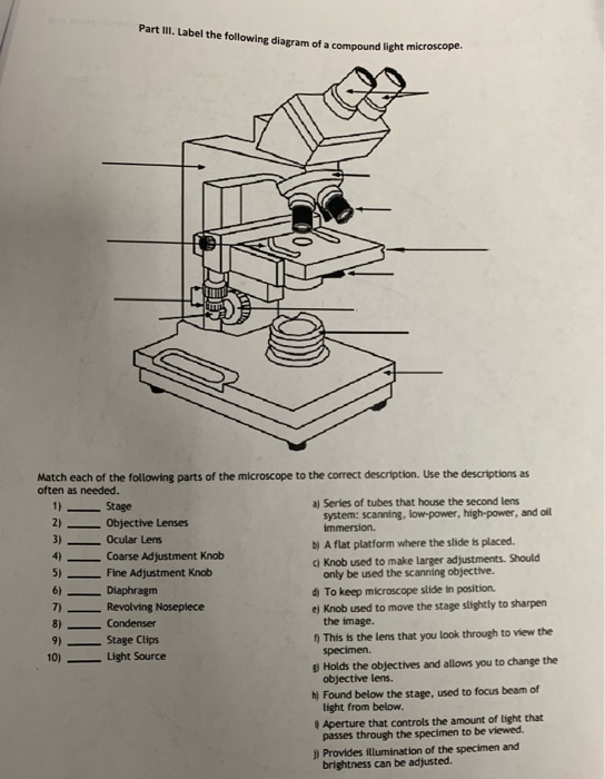
Solved Part III. Label the following diagram of a compound ...
Post a Comment for "45 label microscope diagram"