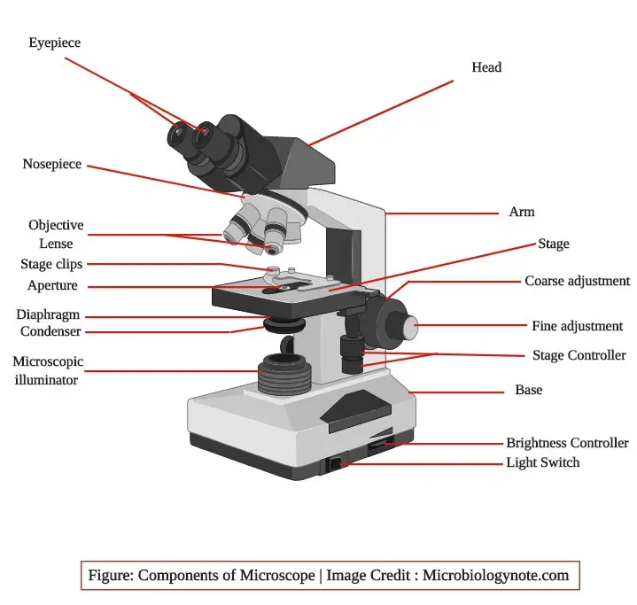
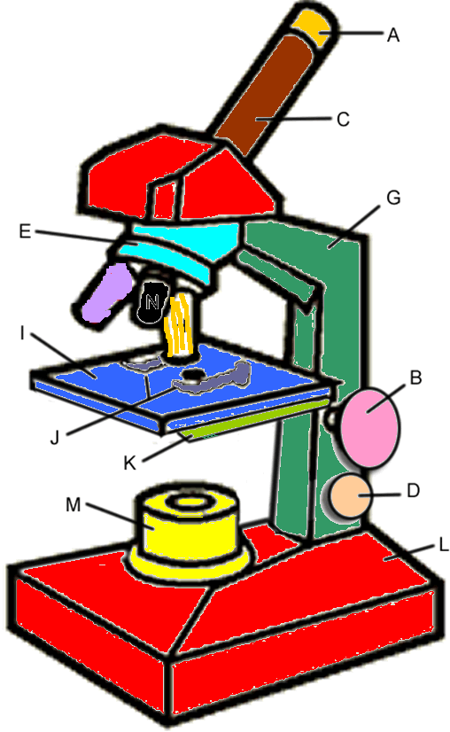
45 label the different parts of the microscope
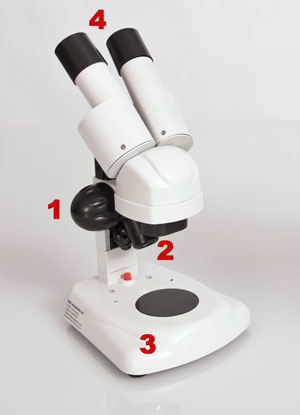
Parts of Stereo Microscope (Dissecting microscope) – labeled … Each light path provides a different angle (usually between 10 and 12 degrees) of viewing in each of our eye. ... Optical parts of a stereo microscope work together to magnify and produce a 3-D image of the specimens. These parts include: ... some eyepieces come with the label “WF” which defines that the eyepiece provides a wide field of ... Virtual Microscope - NCBioNetwork.org Lesson Description BioNetwork’s Virtual Microscope is the first fully interactive 3D scope - it’s a great practice tool to prepare you for working in a science lab. Explore topics on usage, care, terminology and then interact with a fully functional, virtual microscope. When you are ready, challenge your knowledge in the testing section to see what you have learned.
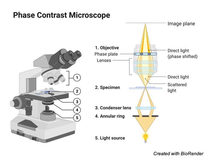
Fluorescence Microscopy - Explanation and Labelled Images Dec 16, 2020 · The Characteristics of a Fluorescence Microscope. The main parts of a fluorescent microscope overlap with the traditional light microscope. However, there are two main features that sets fluorescent microscope apart from the traditional microscope. One is the type of light source and the other is the use of specialized filter elements.

Label the different parts of the microscope
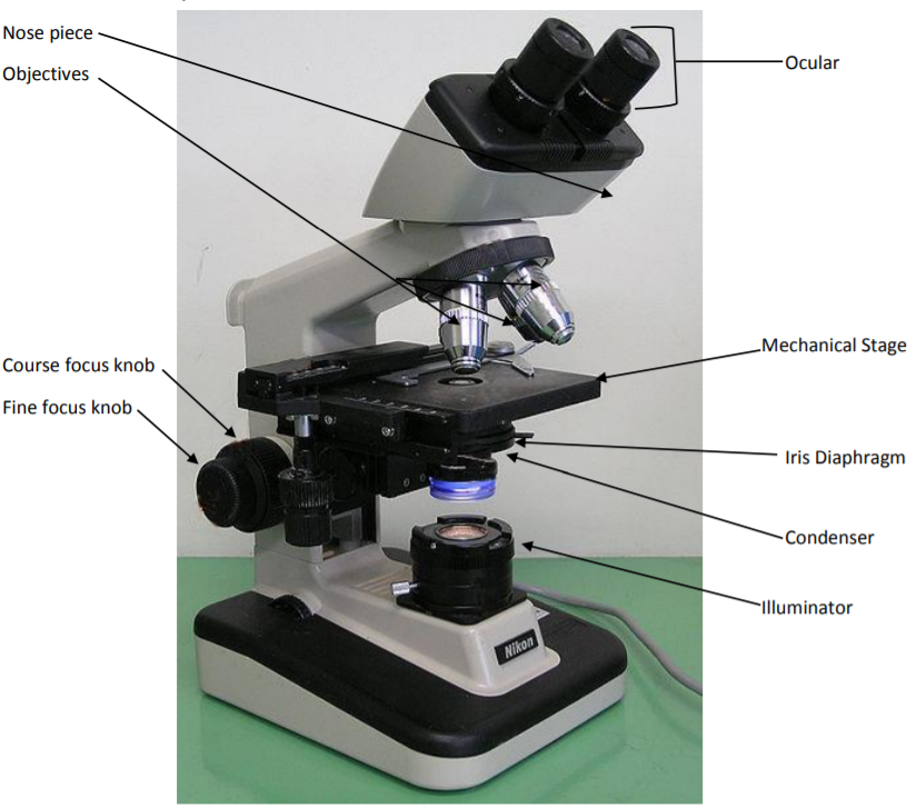
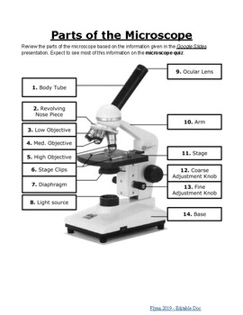
Parts of a microscope with functions and labeled diagram Apr 19, 2022 · Q. Differentiate between a condenser and an Abbe condenser. Ans. Condensers are lenses that are used to collect and focus light from the illuminator into the specimen. They are found under the stage next to the diaphragm of the microscope. They play a major role in ensuring clear sharp images are produced with a high magnification of 400X and above. Label the microscope — Science Learning Hub Jun 08, 2018 · All microscopes share features in common. In this interactive, you can label the different parts of a microscope. Use this with the Microscope parts activity to help students identify and label the main parts of a microscope and then describe their functions.. Drag and drop the text labels onto the microscope diagram. If you want to redo an answer, click on the … Microscope slide - Wikipedia A microscope slide is a thin flat piece of glass, typically 75 by 26 mm (3 by 1 inches) and about 1 mm thick, used to hold objects for examination under a microscope.Typically the object is mounted (secured) on the slide, and then both are inserted together in the microscope for viewing. This arrangement allows several slide-mounted objects to be quickly inserted and …
Label the different parts of the microscope. Virtual Labs: Using the Microscope - GameUp - BrainPOP. In this free online science interactive, students learn the procedures for operating a compound optical light microscope as they would use in a science lab. bVX0-zncj9qJ3G1_r18rkIpQL02X-Oi6tWViR4g4-vwDVmU50WZA-4bRZMjM2TXmc88PAkJ1g0jIembnEbM Microscope, Microscope Parts, Labeled Diagram, and Functions Jan 19, 2022 · Revolving Nosepiece or Turret: Turret is the part of the microscope that holds two or multiple objective lenses and helps to rotate objective lenses and also helps to easily change power. Objective Lenses: Three are 3 or 4 objective lenses on a microscope. The objective lenses almost always consist of 4x, 10x, 40x and 100x powers. The most common eyepiece lens is … What is a Compound Microscope? | Microscope World Blog The Parts & Function of a Compound Microscope A compound microscope is a high power (high magnification) microscope that uses a compound lens system. A compound microscope has multiple lenses: the objective lens (typically 4x, 10x, 40x or 100x) is compounded (multiplied) by the eyepiece lens (typically 10x) to obtain a high magnification of 40x ... Pond Water Under the Microscope Here, students can sketch down what they observe and later label the different parts of the organisms. Conclusion. The primary goal here is for students to observe for themselves the different types of small organisms, which live in the pond and their diversity. Making rough sketches allows them to draw what they see and how they see them.
Microscope slide - Wikipedia A microscope slide is a thin flat piece of glass, typically 75 by 26 mm (3 by 1 inches) and about 1 mm thick, used to hold objects for examination under a microscope.Typically the object is mounted (secured) on the slide, and then both are inserted together in the microscope for viewing. This arrangement allows several slide-mounted objects to be quickly inserted and … Label the microscope — Science Learning Hub Jun 08, 2018 · All microscopes share features in common. In this interactive, you can label the different parts of a microscope. Use this with the Microscope parts activity to help students identify and label the main parts of a microscope and then describe their functions.. Drag and drop the text labels onto the microscope diagram. If you want to redo an answer, click on the … Parts of a microscope with functions and labeled diagram Apr 19, 2022 · Q. Differentiate between a condenser and an Abbe condenser. Ans. Condensers are lenses that are used to collect and focus light from the illuminator into the specimen. They are found under the stage next to the diaphragm of the microscope. They play a major role in ensuring clear sharp images are produced with a high magnification of 400X and above.
Simple Microscope Definition, Magnification, Parts And Uses

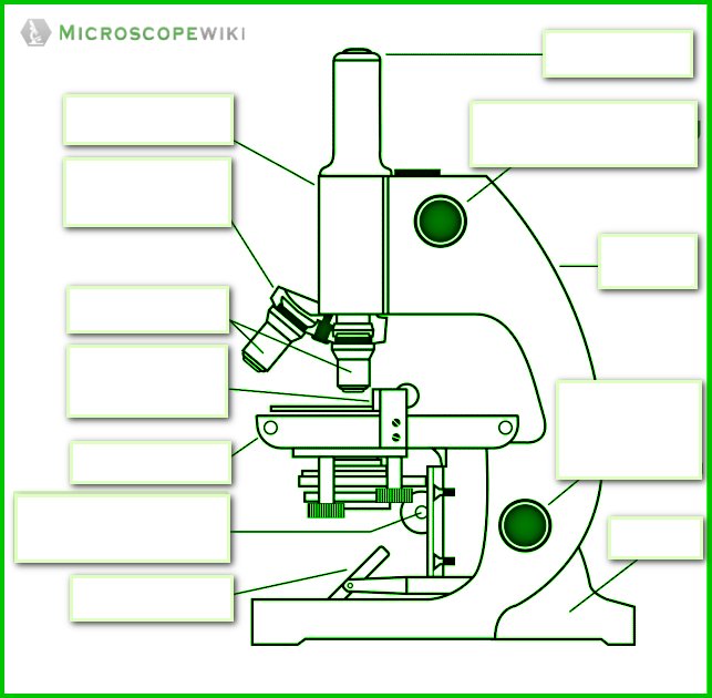
Parts of the microscope interactive worksheet

Microscope Labeling Practice Diagram | Quizlet

Parts of a Microscope - SmartSchool Systems

Christy Peek: November 2015

microscope binokuler led Oregon di Suntik Cantik vitri | Tokopedia

Parts of the Microscope with Labeling (also Free Printouts ...

Label the microscope — Science Learning Hub

Label Microscope Diagram - EnchantedLearning.com
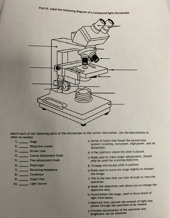
Solved Part III. Label the following diagram of a compound ...

Labeling a Microscope Free Worksheet Pack

Parts of a microscope with functions and labeled diagram
Parts of a Microscope Quiz

This is a common compound microscope Label its parts class 11 ...

Compound Microscope – Diagram (Parts labelled), Principle and ...

Microscope Parts and Functions

Label Microscope Parts - ClipArt Best

Bellwork Why do scientists use Microscopes? - ppt download
4: Microscopy - Biology LibreTexts
Compound and Stereo- microscopes - Microscopes 4 Schools

Parts Of A Microscope Labeling Teaching Resources | TpT
How to see a plant cell under a compound microscope - Quora
Parts of a Microscope

Parts of a Microscope Labeling Activity

The Microscope

Label the microscope — Science Learning Hub
Parts of a Microscope and Their Functions

This is a common compound microscope. Label its parts from A ...
Jual Mikroskop Kid Education 1200x Magnifier science ...

Labeled Microscope Diagram | Microscope parts, Science fair ...

Microscope Maintenance Tips | Science supplies, Microscope ...

Compound Microscope Parts, Functions, and Labeled Diagram ...

Activity 1).docx - Activity #1 THE MICROSCOPE I. A. Label the ...
The Microscope

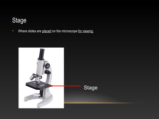
Parts of Stereo Microscope (Dissecting microscope) – labeled ...
Simple Microscope - Diagram (Parts labelled), Principle ...

Microscope Parts and Function | Cell Structure - Quizizz
Microscope, Microscope Parts, Labeled Diagram, and Functions

The Parts of a Microscope (Labeled) Printable Printable (6th ...

The Compound Microscope parts & how they work
Parts of the Microscope Labeling Activity!

Parts of a Compound Microscope and Their Functions
Microscope parts and functions
Label a microscope - Teaching resources
Label Parts Of Microscope - ClipArt Best - ClipArt Best ...
Post a Comment for "45 label the different parts of the microscope"