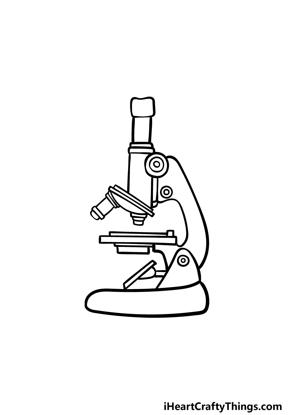
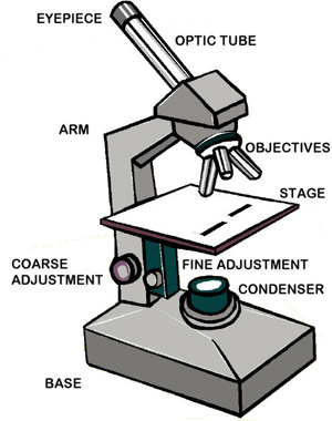
45 draw and label microscope
abcnews.go.com › technologyTechnology and Science News - ABC News Jul 12, 2022 · Get the latest science news and technology news, read tech reviews and more at ABC News. Label Microscope Diagram - EnchantedLearning.com base - this supports the microscope. body tube - the tube that supports the eyepiece. coarse focus adjustment - a knob that makes large adjustments to the focus. diaphragm - an adjustable opening under the stage, allowing different amounts of light onto the stage. eyepiece - where you place your eye.
Microscope Drawing Easy with Label - YouTube Microscope Drawing Easy with Label 886 views Apr 13, 2020 In this video I go over a microscope drawing that is easy with label. There is a blank copy at the end of the video to review on your own....

Draw and label microscope
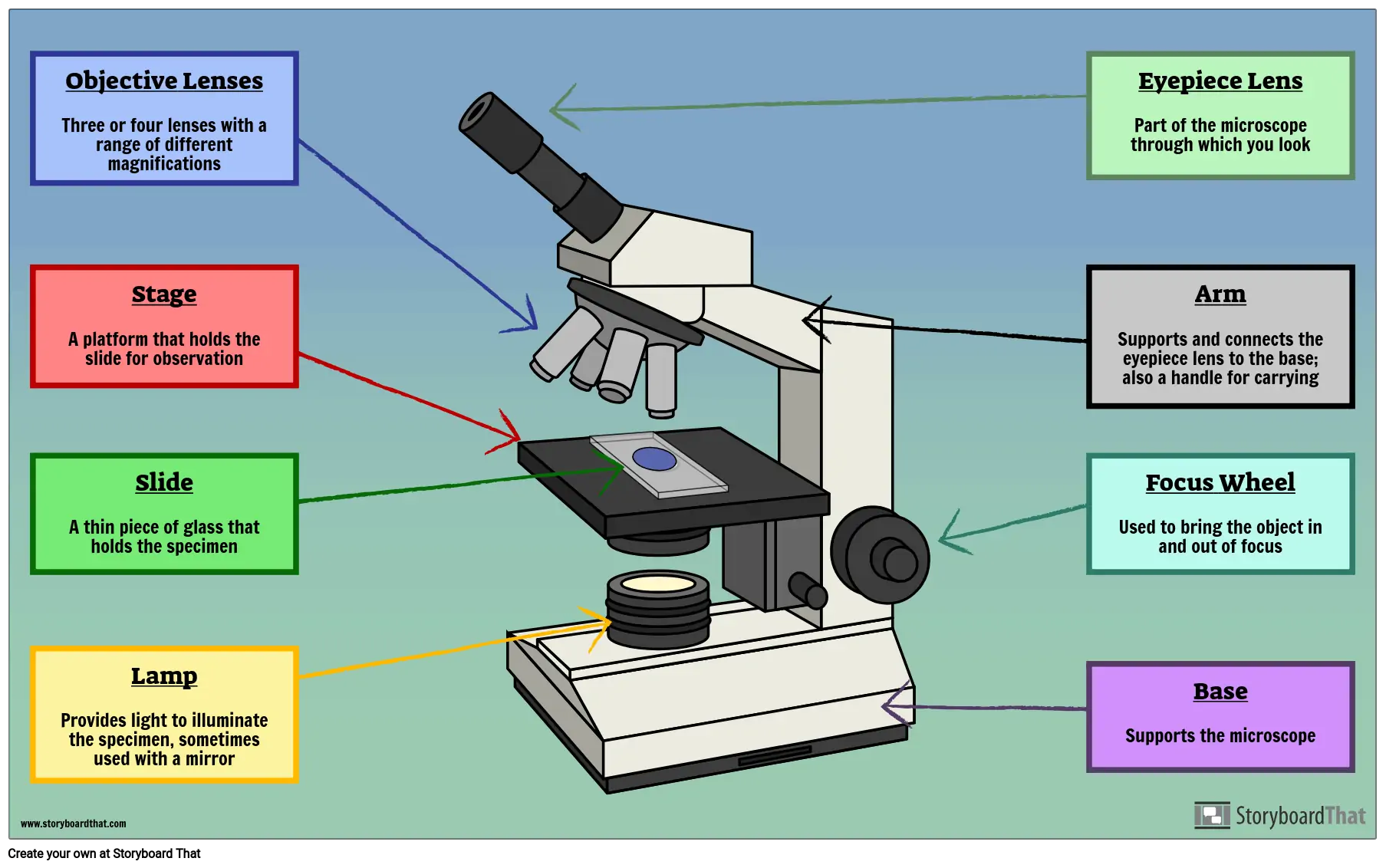
label the microscope worksheet microscope parts worksheet label compound science worksheets diagram printable labeling drawing labels microscopes quiz biology labeled light blank sketch worksheeto. Microscope Labeling Worksheets & Teaching Resources | TpT . microscope magnification labeling calculation. Nervous System - 7 Red Team 7redteam.weebly.com Compound Microscope Parts, Functions, and Labeled Diagram Compound Microscope Definitions for Labels Eyepiece (ocular lens) with or without Pointer: The part that is looked through at the top of the compound microscope. Eyepieces typically have a magnification between 5x & 30x. Monocular or Binocular Head: Structural support that holds & connects the eyepieces to the objective lenses. Microscope Parts, Function, & Labeled Diagram - slidingmotion Microscope Parts Labeled Diagram The principle of the Microscope gives you an exact reason to use it. It works on the 3 principles. Magnification Resolving Power Numerical Aperture. Parts of Microscope Head Base Arm Eyepiece Lens Eyepiece Tube Objective Lenses Nose Piece Adjustment Knobs Stage Aperture Microscopic Illuminator Condenser Lens
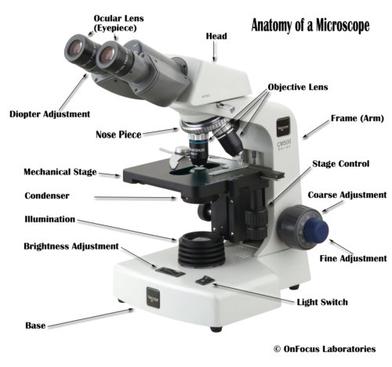
Draw and label microscope. Proper Microscope Drawings and Observations - YouTube This short video discuss the expectations of a microscope observation and drawings and also provides examples of errors to watch out for.Teachers: You can pu... Compound Microscope Parts - Labeled Diagram and their Functions The eyepiece (or ocular lens) is the lens part at the top of a microscope that the viewer looks through. The standard eyepiece has a magnification of 10x. You may exchange with an optional eyepiece ranging from 5x - 30x. [In this figure] The structure inside an eyepiece. The current design of the eyepiece is no longer a single convex lens. microbenotes.com › parts-of-a-microscopeParts of a microscope with functions and labeled diagram Sep 17, 2022 · Figure: Diagram of parts of a microscope. There are three structural parts of the microscope i.e. head, base, and arm. Head – This is also known as the body. It carries the optical parts in the upper part of the microscope. Base – It acts as microscopes support. It also carries microscopic illuminators. Compound Microscope: Definition, Diagram, Parts, Uses, Working ... - BYJUS A compound microscope is defined as. A microscope with a high resolution and uses two sets of lenses providing a 2-dimensional image of the sample. The term compound refers to the usage of more than one lens in the microscope. Also, the compound microscope is one of the types of optical microscopes. The other type of optical microscope is a ...
A Study of the Microscope and its Functions With a Labeled Diagram ... May 21, 2019 - To better understand the structure and function of a microscope, we need to take a look at the labeled microscope diagrams of the compound and electron microscope. These diagrams clearly explain the functioning of the microscopes along with their respective parts. How To Draw A Microscope Step by Step - [12 Easy Phase] In this drawing lesson, we'll show How to draw a Microscope step by step total 12 phase, and it will be easy tutorial COMPOUND LIGHT MICROSCOPE PARTS AND FUNCTIONS .pdf Parts and Functions of a compound light microscope. MAGNIFYING PARTS •EYEPIECE / OCULARS - the lens the viewer looks through to see the magnified specimen. With a magnification of 8x or 10x • DIOPTER (ADJUSTMENT KNOB) - adjusts according to examiner's vision • INTERPUPILLARY DISTANCE (ADJUSTMENT KNOB) - adjusts and adapts to distance between viewer's eyes •OBJECTIVES - the ... A Study of the Microscope and its Functions With a Labeled Diagram The camera present within the microscope captures images to reveal the finer details of the specimen. This microscope can zoom and view the density of a specimen until it is only a micrometer thick and has a magnification ranging between 1,000 - 250,000x on the fluorescent screen. This microscope needs a computer software to yield precise results.
18,701 Microscope drawing Images, Stock Photos & Vectors - Shutterstock 18,701 microscope drawing stock photos, vectors, and illustrations are available royalty-free. See microscope drawing stock video clips Image type Orientation Color People Artists Sort by Popular Science Abstract Designs and Shapes College and University Art Styles Printing, Typography, and Calligraphy microscope line art chemistry laboratory Virtual Microscope - NCBioNetwork.org Lesson Description BioNetwork’s Virtual Microscope is the first fully interactive 3D scope - it’s a great practice tool to prepare you for working in a science lab. Explore topics on usage, care, terminology and then interact with a fully functional, virtual microscope. When you are ready, challenge your knowledge in the testing section to see what you have learned. UD Virtual Compound Microscope - University of Delaware ©University of Delaware. This work is licensed under a Creative Commons Attribution-NonCommercial-NoDerivs 2.5 License.Creative Commons Attribution-NonCommercial-NoDerivs 2.5 License. Microscope Parts and Functions The specimen is placed on the glass and a cover slip is placed over the specimen. This allows the slide to be easily inserted or removed from the microscope. It also allows the specimen to be labeled, transported, and stored without damage. Stage: The flat platform where the slide is placed. Stage clips: Metal clips that hold the slide in place.
Compound Microscope Parts – Labeled Diagram and their ...
Microscope, Microscope Parts, Labeled Diagram, and Functions What is Microscope? A microscope is a laboratory instrument used to examine objects that are too small to be seen by the naked eye. It is derived from Ancient Greek words and composed of mikrós, "small" and skopeîn,"to look" or "see".
Microscope Drawing - How To Draw A Microscope Step By Step
Light Microscope- Definition, Principle, Types, Parts, Labeled Diagram ... A light microscope is a biology laboratory instrument or tool, that uses visible light to detect and magnify very small objects and enlarge them. They use lenses to focus light on the specimen, magnifying it thus producing an image. The specimen is normally placed close to the microscopic lens.

List: Parts of a Microscope and their Function | Pathwooded
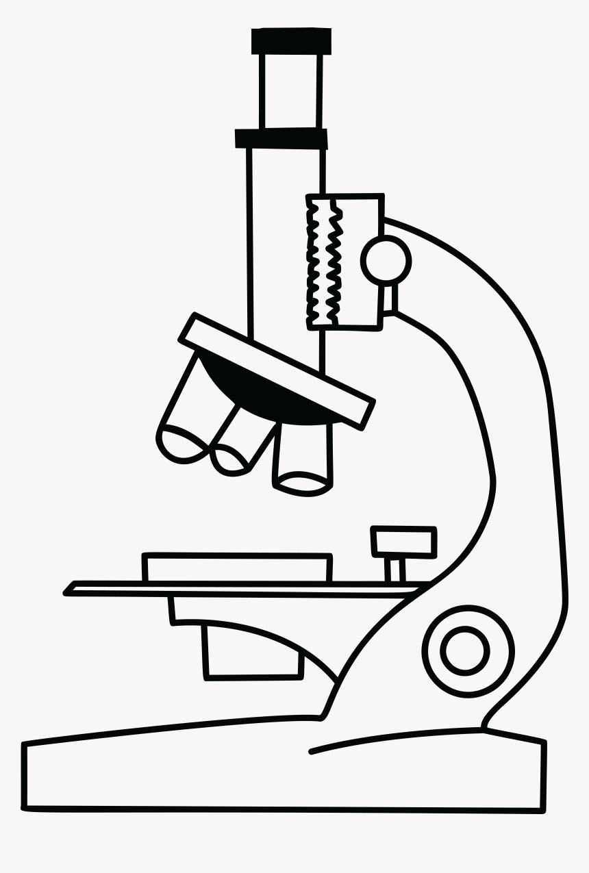
Drawing Of A Microscope And Label - Warehouse of Ideas Here presented 54+ microscope drawing and label images for free to download, print or share. Title Is Informative, Centered, And Larger Than Other Text. How to draw a microscope and label. Compound microscopes have furthered medical research, helped to solve crimes, and they have repeatedly proven invaluable in unlocking the secrets of the.

Microscope Diagram - Label Diagram | Quizlet
Interactive Bacteria Cell Model - CELLS alive Periplasmic Space: This cellular compartment is found only in those bacteria that have both an outer membrane and plasma membrane (e.g. Gram negative bacteria).In the space are enzymes and other proteins that help digest and move nutrients into the cell. Cell Wall: Composed of peptidoglycan (polysaccharides + protein), the cell wall maintains the overall shape of a …

Draw a well labelled diagram of a microscope. - Brainly.in
› microbiology-resource-center › labStains - Microbiology Resource Center - Truckee Meadows ... Wait for capillary action to draw the liquid along the leading edge of the angled slide. Push the angled slide across the surface of the flat slide. Most of the nigrosin should still be left on the original spot. Discard the slide in the disinfectant bucket. Set the stained slide aside to air dry before observing it under oil immersion.
Drawing microscope - Teaching resources
Microscope Drawing: How to Sketch Microscope Slides How to Draw Microscope Slides Organize and orient your field of view: To begin, draw a circle as large as possible with a pencil. An 8.5 x 11-inch piece of paper is good size for beginners. The circle represents what you see through the eyepiece of the microscope. Using thin lines, divide the circle into quarters in order to organize the picture.
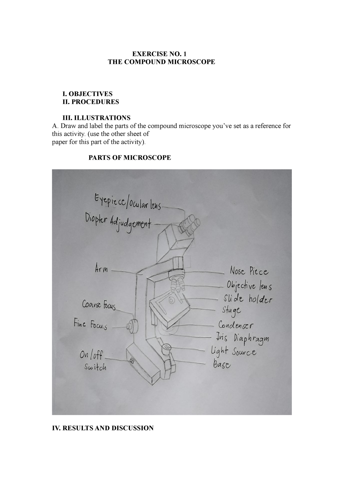
Compound microscopic microbiology - EXERCISE NO. 1 THE ...
Cheek Cell Lab - The Biology Corner Label the nucleus, cytoplasm, and cell membrane of a single cell. Draw your cells to scale. Scanning Low Power: High Power: Analysis. 1. Why is methylene blue necessary? 2. The light microscope used in the lab is not powerful enough to view other organelles in the cheek cell. What parts of the cell were visible. 3. List 2 organelles that were NOT visible but could be …

Simple Microscope - Diagram (Parts labelled), Principle ...
How to Sketch a Microscope Slide - Identifying and Sketching Cell ... First, to represent the microscope field of view, draw a circle on the page - this can be freehand or, if you want to be precise, use a compass. If you are using a graticule slide (a microscope slide with millimeter grid lines), lightly sketch a grid over your circle.
Parts of a Microscope Labeling Activity
Labeling the Parts of the Microscope | Microscope World Resources Labeling the Parts of the Microscope This activity has been designed for use in homes and schools. Each microscope layout (both blank and the version with answers) are available as PDF downloads. You can view a more in-depth review of each part of the microscope here. Download the Label the Parts of the Microscope PDF printable version here.

Microscope - Label - Part 2 Diagram | Quizlet
How To Draw A Microscope 🔬 - YouTube in 2022 | Science diagrams ... Apr 8, 2022 - #microscope #howtodraw #adimushowThis is an easy and simple drawing of microscope .This will teach you how to draw and label a microscope .This is a step-by-...

How to Draw a Microscope - VERY EASY
How To Draw A Microscope - YouTube Today, we're learning how to draw a cool microscope!👩🎨 JOIN OUR ART HUB MEMBERSHIP! VISIT 🎨 VISIT OUR AMAZON ART SUPPLY S...

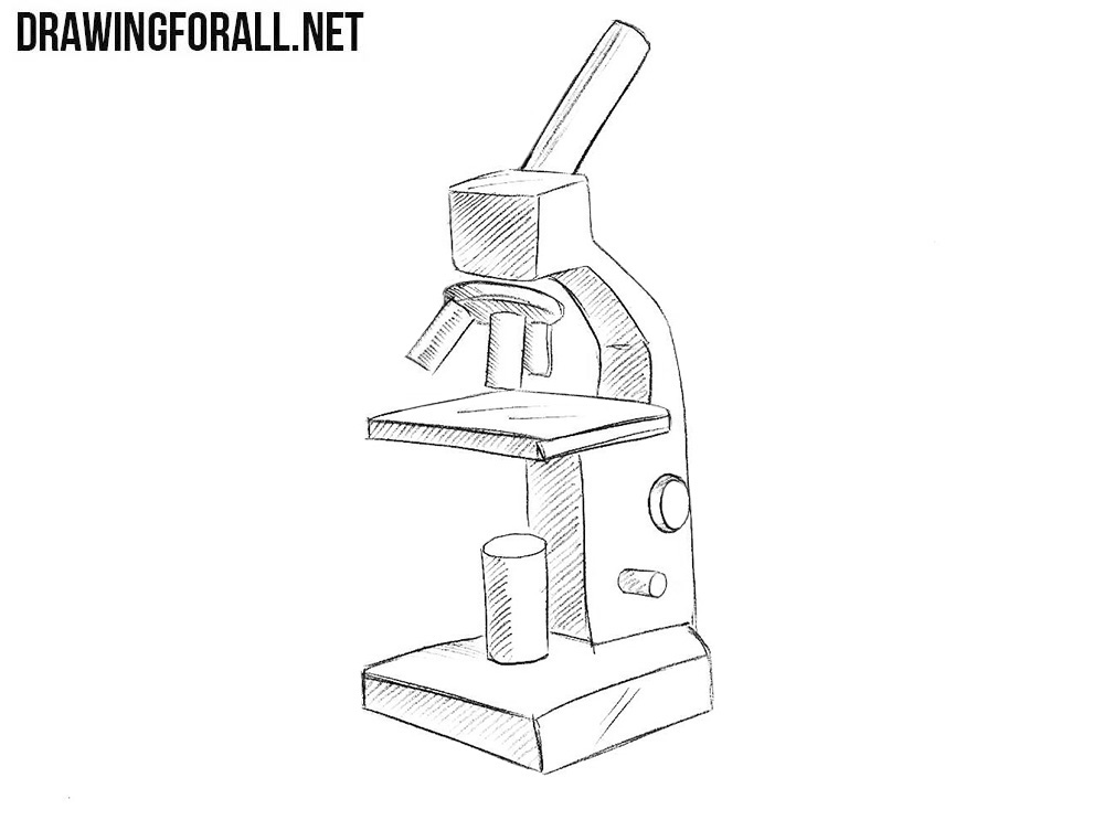
How to draw compound of Microscope easily - step by step
Microscope Drawing Teaching Resources | Teachers Pay Teachers 22. FREE. Word Document File. Use this blank handout as a way for students to record microscope drawings. Aside from the drawig itself, studnts are prompted to title the drawing, include the magnification of the microscope, and give a quick description of what they are viewing. Keywords: Microscope Drawings Lab Biology.
Clip Art Microscope Clipart Black And White With Labels ...
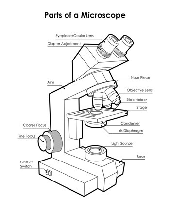
Parts of the Microscope with Labeling (also Free Printouts) Parts of the Microscope with Labeling (also Free Printouts) By Editorial Team March 7, 2022 A microscope is one of the invaluable tools in the laboratory setting. It is used to observe things that cannot be seen by the naked eye. Table of Contents 1. Eyepiece 2. Body tube/Head 3. Turret/Nose piece 4. Objective lenses 5. Knobs (fine and coarse) 6.

Free Microscope Drawing, Download Free Microscope Drawing png ...
How to draw compound of Microscope easily - step by step I will show you " How to draw compound of microscope easily - step by step "Please watch carefully and try this okay.Thanks for watching.....#microscopedrawi...

Course: s4: Biology , Topic: UNIT 3: MICROSCOPY
Simple Microscope - Parts, Functions, Diagram and Labelling A compound microscope is also called a bright field microscope. It can provide magnification by up to 1,000 times. Stereo microscope/dissecting microscope - It can magnify objects by up to 300 times. It is used to visualize opaque objects that cannot be visualized using a compound microscope.

HOw to draw light or compound microscope step by step / Microscope diagram
Compound Microscope- Definition, Labeled Diagram, Principle, Parts, Uses The naked eye can now view the specimen at magnification 400 times greater and so microscopic details are revealed. Alternatively, the magnification of the compound microscope is given by: m = D/ fo * L/fe where, D = Least distance of distinct vision (25 cm) L = Length of the microscope tube fo = Focal length of the objective lens

Simple Microscope Definition, Magnification, Parts And Uses
Microscope Parts, Function, & Labeled Diagram - slidingmotion Microscope Parts Labeled Diagram The principle of the Microscope gives you an exact reason to use it. It works on the 3 principles. Magnification Resolving Power Numerical Aperture. Parts of Microscope Head Base Arm Eyepiece Lens Eyepiece Tube Objective Lenses Nose Piece Adjustment Knobs Stage Aperture Microscopic Illuminator Condenser Lens

Lasec Education | Key parts of a compound microscope and how ...
Compound Microscope Parts, Functions, and Labeled Diagram Compound Microscope Definitions for Labels Eyepiece (ocular lens) with or without Pointer: The part that is looked through at the top of the compound microscope. Eyepieces typically have a magnification between 5x & 30x. Monocular or Binocular Head: Structural support that holds & connects the eyepieces to the objective lenses.

Microscope, Microscope Parts, Labeled Diagram, and Functions
label the microscope worksheet microscope parts worksheet label compound science worksheets diagram printable labeling drawing labels microscopes quiz biology labeled light blank sketch worksheeto. Microscope Labeling Worksheets & Teaching Resources | TpT . microscope magnification labeling calculation. Nervous System - 7 Red Team 7redteam.weebly.com

Label the diagram of the microscope and explain the role of ...

How to draw and label the parts of a microscope? What are at ...
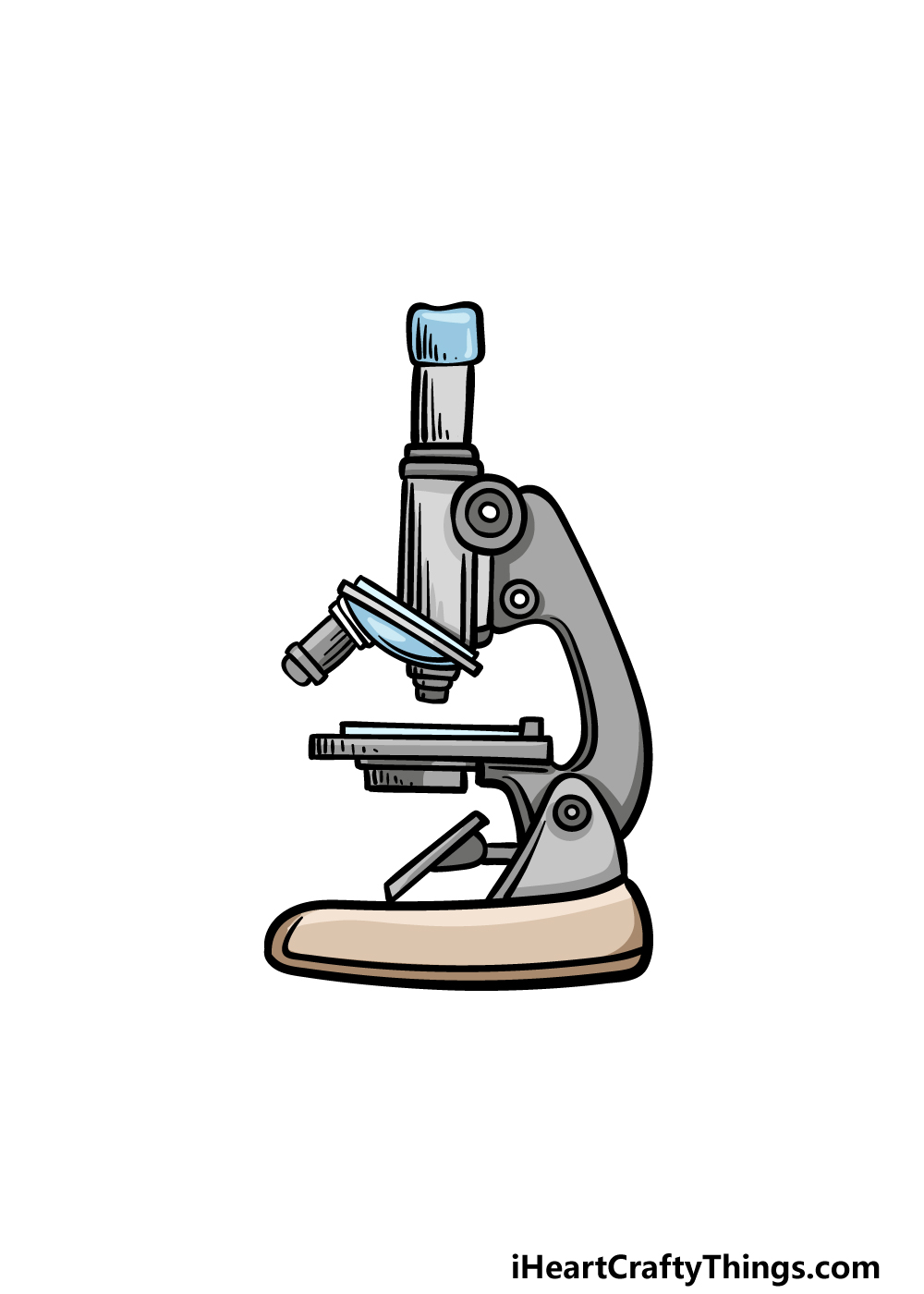
Microscope Drawing - How To Draw A Microscope Step By Step

Living Environment Course

Labeling the Parts of the Microscope | Microscope World Resources

Label Microscope Diagram - EnchantedLearning.com
LAB 1: Scientific Method/Tools of Scientific Inquiry

Compound Microscope: Parts of Compound Microscope

MICROSCOPE Labeling - Part - 3

microscope | Types, Parts, History, Diagram, & Facts | Britannica

Parts of a microscope with functions and labeled diagram
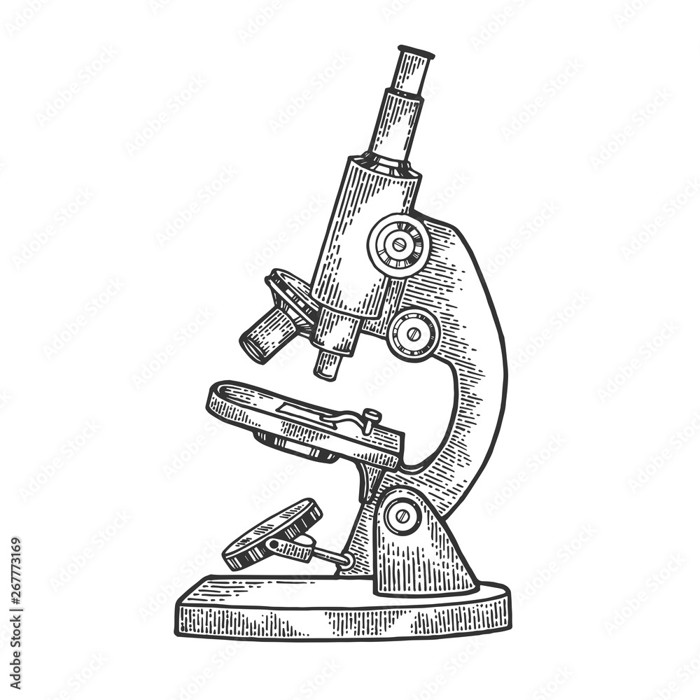
Old Microscope sketch engraving vector illustration. Scratch ...

Diagram of a Compound Microscope

Parts of a microscope with functions and labeled diagram
How to Draw a Microscope Easy
Comparing and Contrasting the Different Parts of the Microscope

Microscope - diagram Tom Butler | Science skills, Science ...
Labeled Microscope Diagram – Tim's Printables
1.2: Microscopes - Biology LibreTexts

How to draw Microscope diagram for beginners - step by step
PRACTICAL BOOKLET - BIOLOGY4ISC
SFCC Biology 160 - Documents

Microscope Parts & Functions - AmScope

Parts of the Microscope with Labeling (also Free Printouts ...
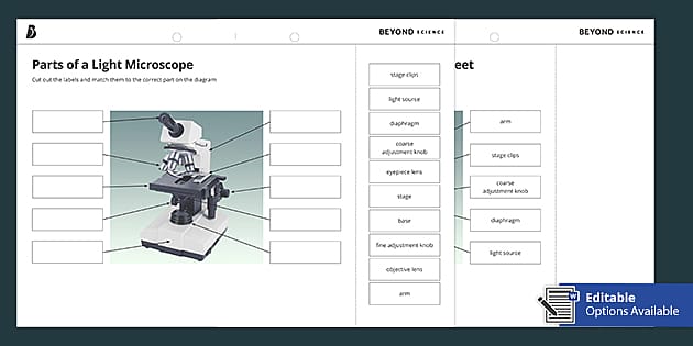
Parts of a Light Microscope Cut and Stick Worksheet - Twinkl
Microscope Parts and Functions
Post a Comment for "45 draw and label microscope"